WHAT IS PATELLA DISLOCATION? Patella dislocation is among the most common injuries on human body. It often occurs in teenagers and young adults, after either a contact or no contact injury. Patella dislocation mostly occurs when the knee is almost in full extension. Nearly always the patella is being dislocated towards the lateral part of the knee. Patella relocation could easily be succeeded by extending the knee and slightly pushing it back into the trochlea. Thus the patella, which must be centered in the femoral trochlea, regains its normal position.
HOW DO YOU TREAT PATELLA DISLOCATION? When a patella dislocation occurs for the first time, an assesment by an expertised Orthopedic Surgeon must be obtained. He will ask for specific medical history(mechanism of injury etc.), special x-rays, MRIs and probably a C/T scan. The injury is initially treated conservatively, if the "geometry" and biomechanical function of the patellofemoral joint is normal and there is no detached osteochondral fragment from the patella or the lateral femoral condyle. If the biomechanics of the joint is abnormal, it is considered almost certain that the patient will experience a patella dislocation again, and usually surgery treatment is recommended. The expertised Orthopedical Surgeon must recognise properly all causes and complete pathology of the injury, in order to choose the suitable plan of treatment for each patient! Causes of recurrent patella dislocation are multiple and may include: patella alta, tight lateral retinaculum, medial patellofemoral ligament (MPFL) rupture, trochlea dysplasia (Dejour's classification is commonly used), dysplastic patella, femoral anteversion, tortional tibia deformity, increased Q-angled, valgus knees etc.
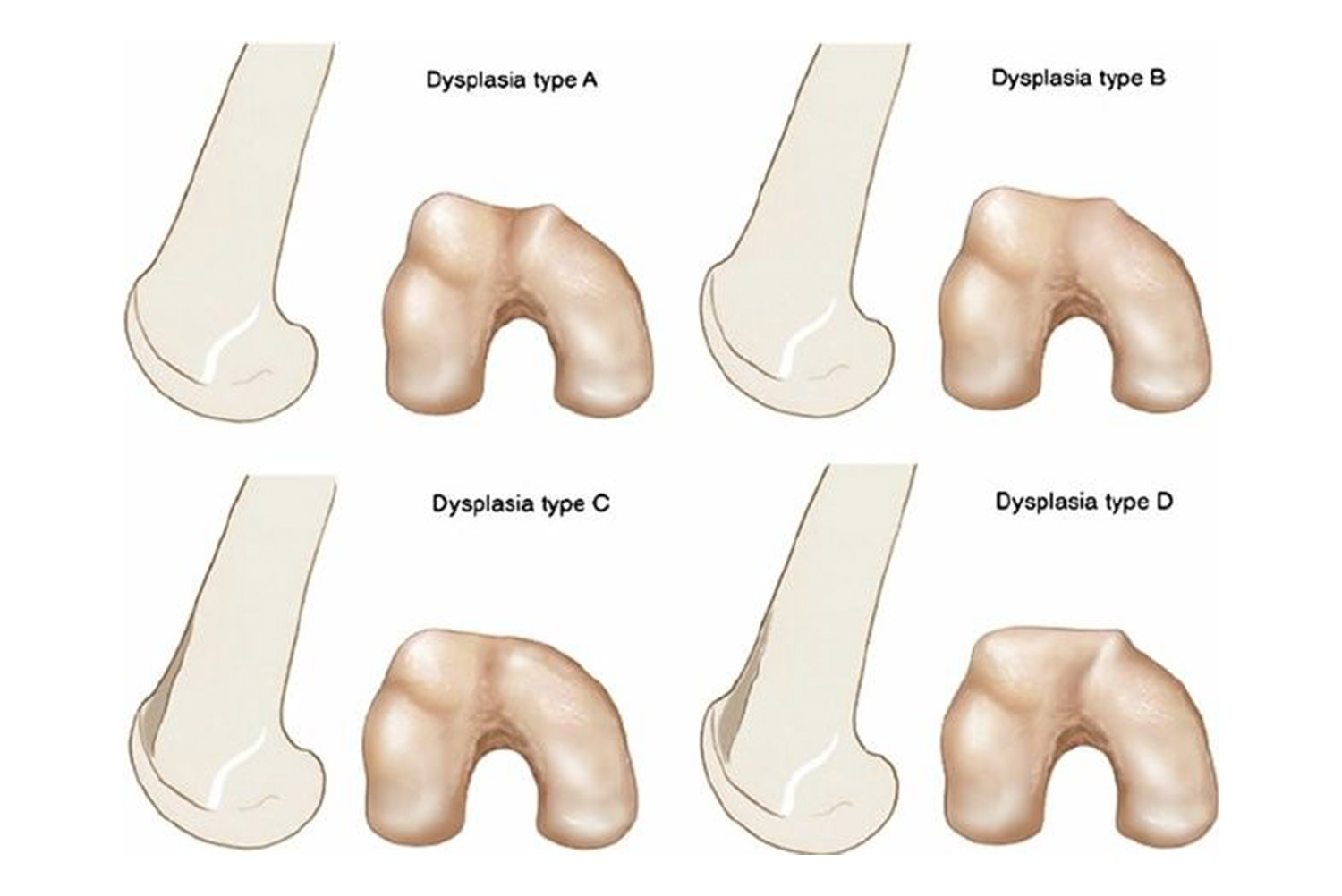
DESCRIPTION OF TROCHLEA DYSPLASIA One area of the knee that is not often discussed is the trochlea. It is the edge of the femur, underneath the patella. This area is completely covered by cartilage and has a “v-shape”, in order to fully harmonize with the relative patella, which "slips" on it. The lateral wall of the trochlea is higher and larger than the medial, with a ratio of 2/3 to 1/3. Thus, the patella can move effortlessly between the walls of the trochlea, which keep it stabilized. If the trochlea is dysplastic, the patella is unstable and it is pushed out of the trochlea. This is the cause of patella dislocation! Patella dislocation is a very severe condition, since patella is considered the bone that transduces and multiplies the strength of the quadriceps to the shinbone. It is widely considered to be the most significant bone for the proper function of the knee! Trochlea dysplasia is a condition that the trochlea appears relatively shallow, flat or in the shape of a "dome". Dejour was the first to classify trochlea dysplasia into four grades (A, B, C and D).
TROCHLEA DYSPLASIA SYMPTOMS
- Pain at the knee and the patella
- High rates of patella dislocation and recurrent instability
WHAT KIND OF RADIOLOGICAL EXAMINATION IS REQUIRED? The expertised Orthopedic Surgeon should take a full and thorough patient’s medical history and ask for specific x-rays, MRIs and usually a C/T scan. Trochlea dysplasia is classified as A, B, C, D (see. Photo 1).
TROCHLEA DYSPLASIA TREATMENT The treatment of trochlea dysplasia is usually surgery and is called trochleoplasty! An experienced orthopedic will restore normal anatomy of the trochlea so that the patella can "slip" properly when bending the knee. Thus, through trochleoplasty, the proper biomechanics of the knee is restored. The probability of recurrence of the patella dislocation decreases from 70% to 1%! Other operations, such as lateral release, tibia tubercle transfer and/or medial patellofemoral ligament (MPFL) reconstruction, are very likely to be performed at the same time as trochleoplasty. It is therefore understood that each patient is unique and each requires attention and individualized treatment by the specialist, an Orthopedic Surgeon specialized in the field of Sports Injuries and Sports Medicine.
 Photo 1. Trochlea Classificationς according to Dejour
 Photo 2. Preoperative Computed Tomography – Trochlea Dysplasia C (Dejour classification)
 Photo 3. Immediate postoperative CT scan - Complete restoration of the normal anatomy of the trochlea.